


New funding accelerates development of smart lung sensor for heart failure and respiratory illness
A new device being developed at the University of Washington could transform how clinicians diagnose and monitor respiratory illness. The Lung Ultrasound Sensor (LUSS) is the work of Dr. Adeyinka Adedipe and a multidisciplinary team, including Drs. Ross Kessler, Kennedy Hall, Tatiana Khokhlova, and Michael Bailey.
The idea emerged in the early months of the COVID-19 pandemic, as patients frequently arrived with severe shortness of breath but with unclear underlying causes.
“We needed to understand how their lungs were functioning,” Dr. Adedipe said. “Ultrasound was a valuable imaging tool, but at the time it required providers to be in the room with patients, which carried significant risks.”
The team set out to design a device that could record lung function using ultrasound without a trained operator. Early funding allowed them to develop a portable, hands-free, non-imaging sensor that detects fluid buildup in the lungs, a hallmark of pulmonary edema. The final version will show results as green, yellow, or red alerts.
“We intend to make it similar to an EKG sticker, but for the lungs,” Adedipe explained. “It shows when lungs are clear and when immediate action is needed because of edema.”
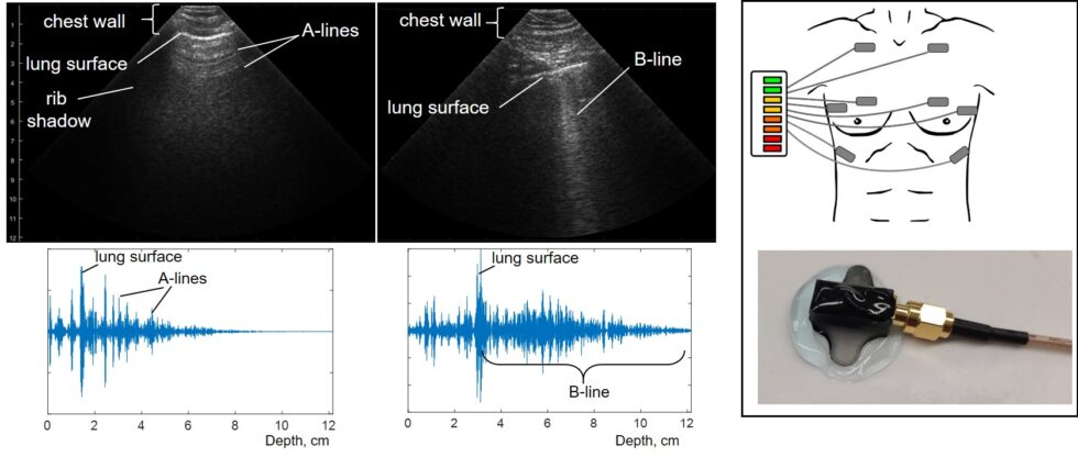
The project recently received UW Innovation Gap Fund support, which backs proof-of-concept and business development for high-potential innovations. This funding will allow the team to refine the prototype and collaborate with engineers at UC San Diego to develop a flexible adhesive patch that wirelessly transmits ultrasound data.
Looking ahead, the researchers aim to bring the technology to market, starting with emergency medical services and prehospital care.
“Right now, EMS providers rely on stethoscopes or pulse oximeters, which aren’t particularly accurate for detecting pulmonary edema,” Adedipe said. “This technology could fundamentally change how we diagnose the condition, track treatment success, and safely discharge patients sooner.”