

Wearable lung sensor could help detect heart failure earlier
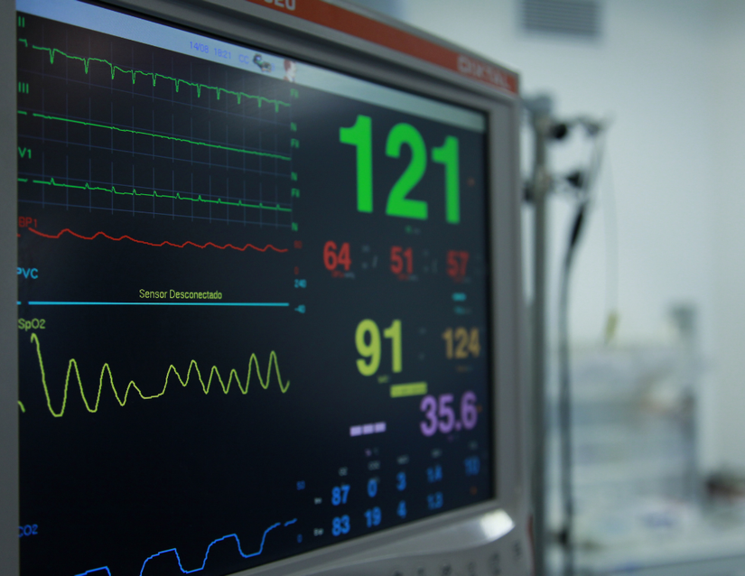
A University of Washington research team is entering the next phase of developing a wearable lung ultrasound sensor that could transform how clinicians detect and monitor fluid buildup in the lungs.

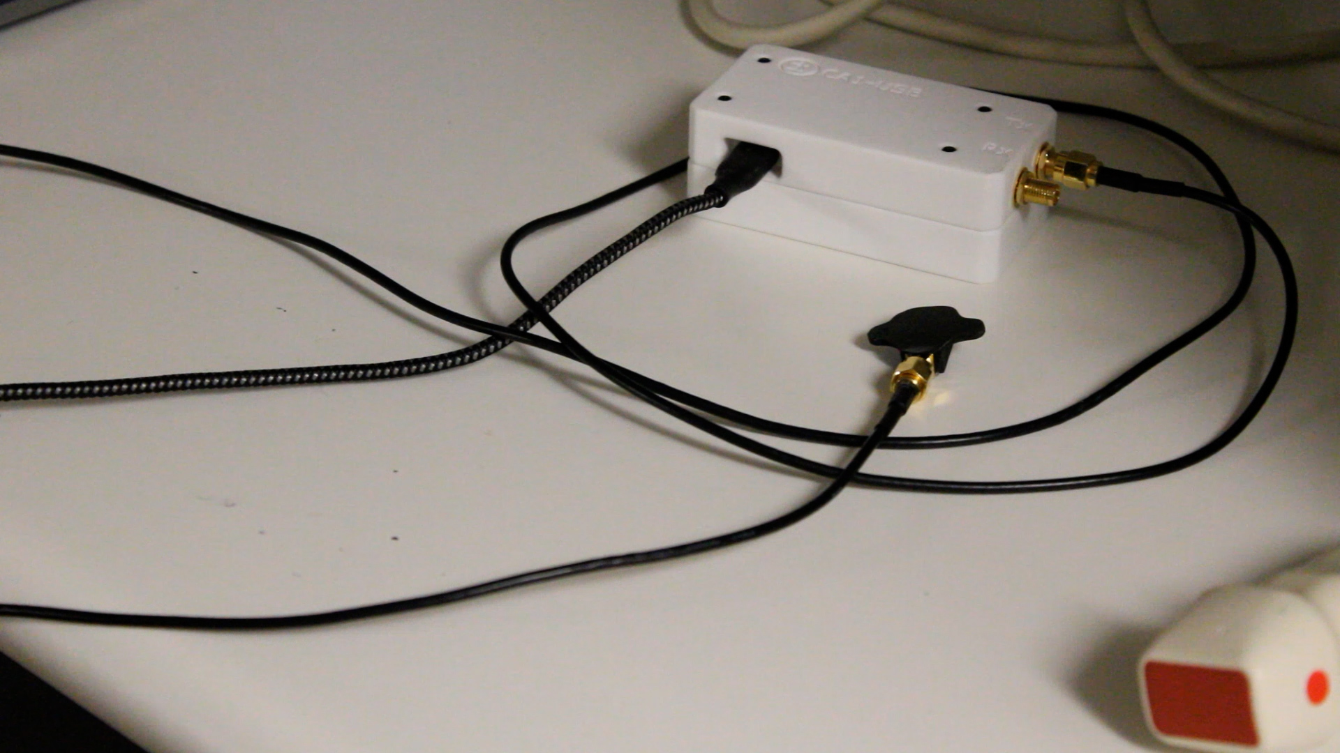
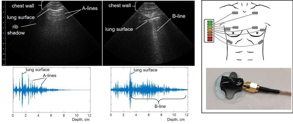
Led by emergency medicine physician Dr. Adeyinka Adedipe, the multidisciplinary team is developing a lightweight, wearable Lung Ultrasound Sensor (LUSS) to detect worsening pulmonary edema. Often caused by heart failure, pulmonary edema occurs when fluid builds up in the lungs, making it increasingly difficult to breathe. The collaboration includes researchers from the UW Department of Emergency Medicine and the Center for Industrial & Medical Ultrasound (CIMU), including Drs. Tatiana Khokhlova and Michael Bailey of CIMU and Drs. Ross Kessler and Kennedy Hall of the Department of Emergency Medicine.
The project originated during the COVID-19 pandemic, when emergency departments were overwhelmed with patients experiencing severe respiratory illness. Lung ultrasound quickly became an important tool for evaluating how well patients' lungs were functioning, but it required clinicians to repeatedly enter patient rooms to perform exams.
"We were able to capture images of patients with respiratory distress who had profound lung edema. And with that, we determined that this could have broad applicability for other patients with other lung conditions," Adedipe said. "At the time it required providers to be in the room with patients, which carried significant risks."
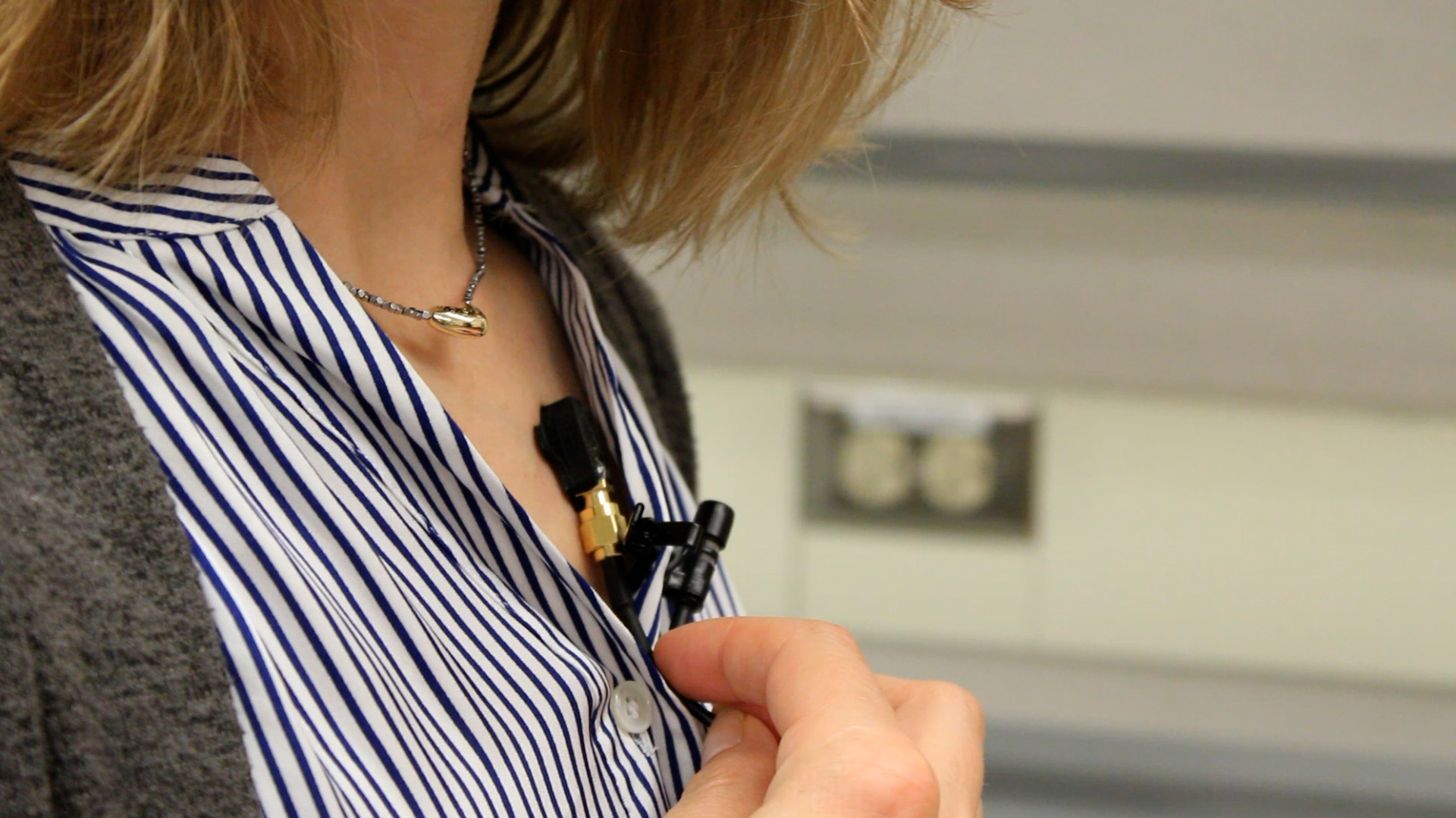
That experience sparked a collaboration between Emergency Medicine and the Center for Industrial & Medical Ultrasound to develop a hands-free device that researchers hope can one day be used by first responders, clinicians and patients at home.
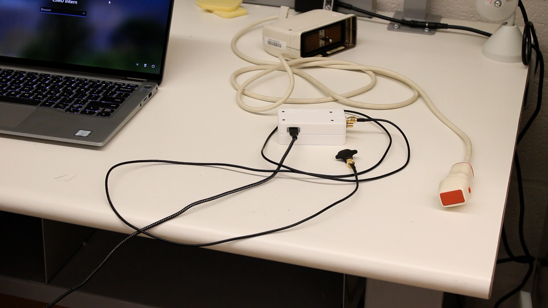
"Right now, EMS providers rely on stethoscopes or pulse oximeters, which aren't particularly accurate for detecting pulmonary edema," Adedipe said.
The goal is to detect fluid buildup before severe symptoms develop. Using a simple green, yellow and red indicator, the wearable sensor could quickly show whether fluid levels are normal, rising or require urgent attention, giving patients and clinicians information they can use to guide treatment, potentially avoid unnecessary hospital visits and improve recovery after discharge.
"It's peace of mind," said Tatiana Khokhlova, PhD, co-investigator on the project. "It will save you those scary moments of 'I can't breathe.'"
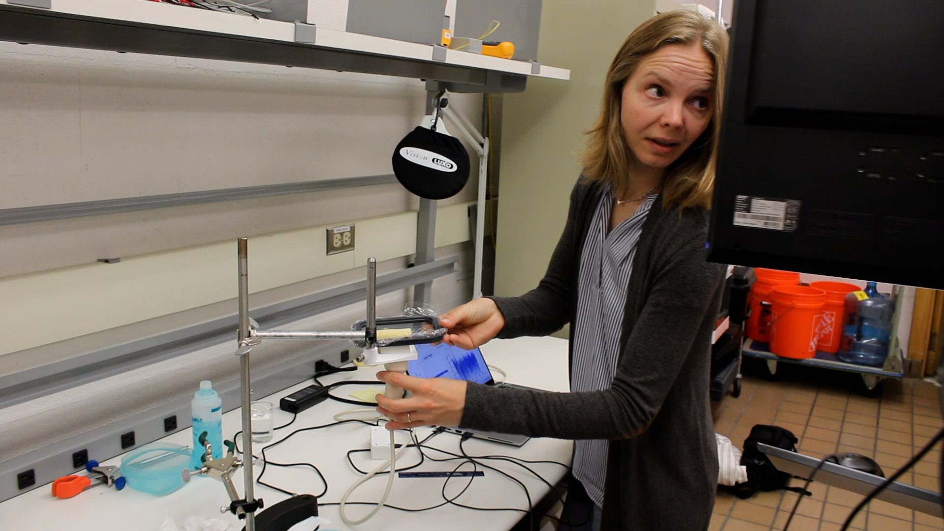
Following an initial clinical study comparing the wearable sensor with standard lung ultrasound, the project recently received support from the University of Washington Royalty Research Fund to launch pilot clinical testing of the latest prototype at UW Medical Center. During the study, researchers will continue refining the device to make it smaller, wireless and more comfortable for patients while evaluating its performance in a clinical setting.
Additional support from UW CoMotion's Gap Fund is helping move the technology toward commercialization. The team is pursuing a licensing agreement through CoMotion and laying the groundwork for a spinout company to manufacture the device and prepare for larger, multi-site clinical trials.
While heart failure remains the primary focus, researchers believe the technology could eventually be used to monitor pneumonia, acute respiratory distress syndrome (ARDS) and lung inflammation in firefighters, military personnel and others exposed to smoke or airborne particulates. If development continues as planned, the team hopes the technology will advance to larger clinical trials over the next year.
“This technology could fundamentally change how we diagnose the condition, track treatment success and safely discharge patients sooner,” said Adedipe.